
Primary Canines
- There are four primary canine teeth, two in eachdental arch.
- These primary canines differ from the outline of their permanent successors in the following ways:
- The crown of the primary maxillary canine (C and H) has relatively longer and sharper cusp than that of its permanent successor on eruption.
- The mesial and distal outlines of the primary maxillary canine are rounder.
Various views of a primary maxillary canine. (From Bath-Balogh MB, Fehrenbach MJ: Illustrated dental embryology, histology, and anatomy, ed 2, Philadelphia, 2005, Saunders.)

You may be interested
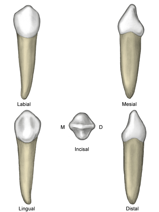
Primary Mandibular Canines
- The primary mandibular canine (M and R) resembles the primary maxillary canine.
- This tooth is much smaller labiolingually.
- The distal cusp slope is much longer than the mesial cusp slope.
- The lingual surface of the primary mandibular canineis marked by a shallow lingual fossa.
- The primary mandibular canine (M and R) resembles the primary maxillary canine, although some dimensions are different. This tooth is much smaller labiolingually.
Various views of a primary mandibular canine. (From Bath-Balogh MB, Fehrenbach MJ: Illustrated dental embryology, histology, and anatomy, ed 2, Philadelphia, 2005, Saunders.)
Primary Molars
- The crown of the primary maxillary first molar(B and I) does not resemble any other crown of either dentition.
- The height of contour on the buccal surface is at the cervical third of the tooth and on the lingual side is at the middle third.
- The primary maxillary molars have three roots, which are thinner and have greater flare than do those of the permanent maxillary first molar.
- The lingual root is the longest and most divergent.
Various views of a primary maxillary first molar. (From Bath-Balogh MB, Fehrenbach MJ: Illustrated dental embryology, histology, and anatomy, ed 2, Philadelphia, 2005, Saunders.)
Primary Maxillary Second Molars
- The primary maxillary second molar (A and J) is larger than the primary maxillary first molar.
- This tooth most closely resembles the permanent maxillary first molar but is smaller in all dimensions.
- The second molar usually has a cusp of Carabelli, the minor fifth cusp.
Various views of a primary maxillary second molar. (From Bath-Balogh MB, Fehrenbach MJ: Illustrated dental embryology, histology, and anatomy, ed 2, Philadelphia, 2005, Saunders.)

Primary Mandibular First Molars
- The crown of the primary mandibular first molar (L and S) is unlike any other tooth of either dentition.
- The height of contour on the buccal surface is at the cervical third of the tooth and on the lingual side is at the middle third.
- The primary mandibular first molar has four cusps; the mesial cusps are larger.
- The tooth has two roots, which are positioned similarly to those of other primary and permanent mandibular molars.
Various views of a primary mandibular first molar. (From Bath-Balogh MB, Fehrenbach MJ: Illustrated dental embryology, histology, and anatomy, ed 2, Philadelphia, 2005, Saunders.)

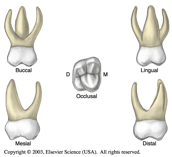
Primary Mandibular Second Molars
- The primary mandibular second molar (K and T) is larger than the primary mandibular first molar.
- It has five cusps; the second molar most closely resembles the permanent mandibular first molar.
- The three buccal cusps are nearly equal in size.
- The primary mandibular second molar has an overall oval occlusal shape.
Various views of a primary mandibular second molar. (From Bath-Balogh MB, Fehrenbach MJ: Illustrated dental embryology, histology, and anatomy, ed 2, Philadelphia, 2005, Saunders.)

Source: https://t-tees.com
Category: WHICH